Regional flux analysis of longitudinal atrophy in Alzheimer's disease
Résumé
Background. The longitudinal analysis of the brain morphology in Alzheimer's disease (AD) is fundamental for discovering and quantifying the dynamics of the pathology. We can broadly identify two main paradigms for the analysis of time series of structural magnetic resonance images (MRIs): hypothesis-free and regional analysis. In the former case, the longitudinal atrophy is modeled at fine scales on the whole brain such as in the voxel/tensor based morphometry and cortical thickness analysis.These methods are useful for exploratory purposes, but usually lack robustness for a reliable quantification of the changes at the subject level. On the other hand, the regional analysis identifies volume changes in preliminary segmented regions. It is however limited to previously defined regions of interest, and therefore it might fail to detect the complex and spread pattern of changes which is likely to underlie the evolution of the pathology. In this study we propose the regional flux analysis, a new approach for the study of the brain longitudinal changes. The aim of regional flux analysis is twofold: consistently unify hypothesis-free and regional approaches to 1) reliably discovery the dynamics of brain morphological changes, and 2) at the same time provide statistically powered measures of longitudinal atrophy. Methods. We encode the morphological differences of follow-up images by longitudinal deformations estimated by non-linear image registration. We compute the scalar pressure potential associated to the non-linear deformations, and we identify the regions of maximal apparent volume change by the loci of extremal pressure. Maximum pressure points identify significant areas of volume loss (deformation sinks), while minimum pressure points identify significant areas of volume gain (deformation sources). We build an atlas of probabilistic regions of group-wise significant sources and sinks of longitudinal atrophy, which is used as reference for quantifying the volume changes of given patients as the flux of the longitudinal deformation across these regions. We tested our method on the discovery and measurement of the yearly longitudinal atrophy of 200 healthy controls, 150 subjects with mild congnitive impairment (MCI) and 142 AD patients. For each subject, baseline and 1-year images were non-linearly registered with the LCC-logDemons algorithm. The probabilistic atlas was estimated from a subset of longitudinal deformations estimated for 20 AD patients, and the resulting regions were used for the quantification of the longitudinal atrophy in the remaining subjects. Statistical power of the resulting measures was assessed by sample size analysis. Results. The estimated probabilistic atlas was composed by 44 and 18 regions of respectively deformation sink and sources. The sink regions of apparent volume loss mapped to grey/withe matter regions, and included hippocampi (bilateral), temporal areas (Sup,Mid and Inf temporal gyrus), Insula and Parahippocampal gyrus. The source regions of apparent volume gain were localized exclusively in CSF areas, among the which Posterior, Anterior and Temporal horns of the ventricles. Longitudinal atrophy measured in hippocampi, temporal regions, and temporal horn of the ventricles was the most discriminative between controls and respectively MCI and AD. Based on the whole set of longitudinal atrophy measurements, sample size analysis required 243 (95% CI: 151,441) and 556 (95% CI: 244,1273) subjects per arm when considering respectively AD and MCI for a randomized two-arm placebo controlled clinical trial for detecting 25% atrophy reduction by controlling for normal aging (80% power, p=0.05). On the head-to-head comparison, the proposed flux analysis outperformed in terms of reduced sample size previously validated quantification methods based on longitudinal hippocampal volumetry. Conclusions. Regional flux analysis of deformations is a novel approach to deformation based morphometry which combines the flexibility of voxel based methods (like tensor based morphometry) with the robustness of segmentation based methods for the quantification of longitudinal atrophy. We showed that regional flux analysis enables a fully automated and powered analysis of longitudinal atrophy in AD, and favorably compares with validated methods for the regional quantification of longitudinal atrophy. Flux analysis thus represents a promising candidate for detecting and robustly quantifying potential drugs effects in clinical trials.
Domaines
Imagerie médicale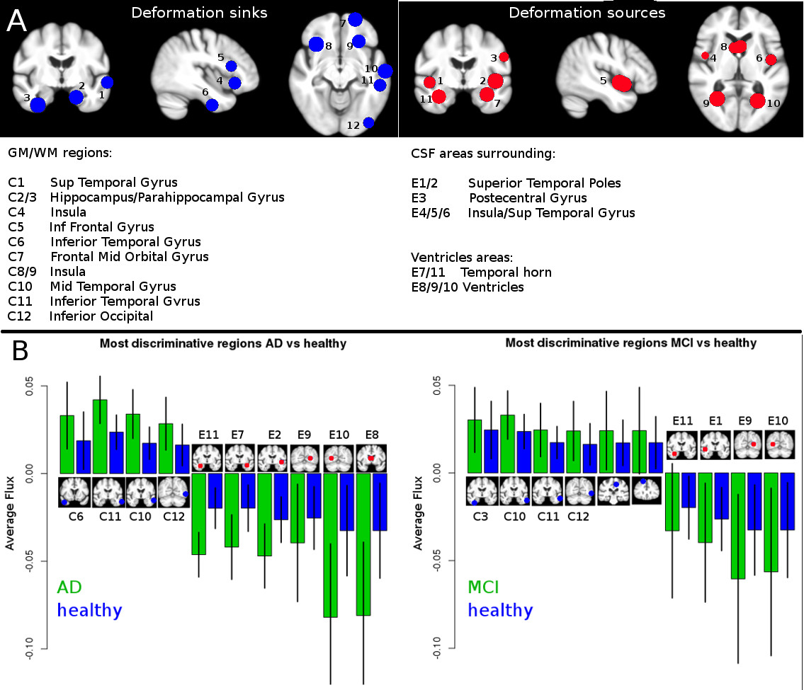
Format : Figure, Image
Origine : Fichiers produits par l'(les) auteur(s)
Origine : Fichiers produits par l'(les) auteur(s)
Format : Papier court
Origine : Fichiers produits par l'(les) auteur(s)
Origine : Fichiers produits par l'(les) auteur(s)
Origine : Fichiers produits par l'(les) auteur(s)